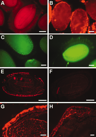
Figure 3. Immunofluorescence of wild-type (Col-2) and mum4-1 mucilage with antipectin antibodies. A, Immersion immunofluorescence of whole wild-type seeds, labeled with -PGA/RGI primary and anti-rabbit Alexa 594 secondary antibody. Note autofluorescence of seed itself and stain of mucilage capsule surrounding the seed. B, Immersion immunofluorescence of whole mum4-1 seed, labeled with -PGA/RGI after shaking in ammonium oxalate. The stained mucilage capsule is thin compared with wild type. C and D, Immersion immunofluorescence of whole wild-type seeds, labeled with JIM7 as primary antibody, followed by anti-rat fluorescein isothiocyanate secondary antibody. C, Seed treated as in A with no shaking pretreatment. No stain of mucilage capsule. D, Seed pretreated by imbibition of extensive shaking in water. Stain of mucilage capsule is apparent. E to H, Immunofluorescence of sections of 9-DPA seeds incubated with -PGA/RGI or serum control. E, Wild-type seed incubated with -PGA/RGI antibody, epidermal cells are labeled on seed surface. F, Wild-type seed incubated with nonimmune normal rabbit serum control, no staining of seed epidermis, same photographic conditions as E. G, Direct comparison of wild-type seed and H mum4-1 seed show reduced -PGA/RGI label in mum4 mutant. Scale bars: A to F = 100 µm and G and H = 50 µm.